SUBJECT TEACHER: C.J. ANGELA J. PINUGU

Study of the organized effort in the management of radiological facility to ensure consistent production of high standard images with minimum exposure to patient and personnel. It discusses quality management and quality assurance systems used for all radiological facilities. Procedures for maintenance are also included. It is a presentation of differentiate test tools to ensure quality output of radiographs and images produced using the different modalities. The lecture component presents and discusses the theories underlying the concepts and procedures for quality assurance and quality control. Activities such film analysis and regular checking monitoring are also incorporated in the discussion of the produced quality images.
This is the study of the general foundations of positioning technique to
obtain radiographic demonstration of anatomical structures of interest as well
as specialized radiographic examinations of the different structures and organs
without contrast media. This includes anatomic and radiographic positioning
terms, source image receptor distance and tube film alignment, positioning
principles, radiographic landmarks, exposure techniques, structures
demonstrated and evaluation criteria of examinations of the different organs.
Clinical competency is very essential and is done through positioning
demonstration and return demonstrations as part of their laboratory procedures.
-KRISTINE KATE S. PAMITTAN, RRT (SUBJECT TEACHER)
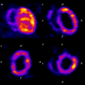
Radiologic knowledge and best practices are constantly evolving. Changes in research methods, professional practices, or medical treatment may become necessary or appropriate as new research and experience broaden our understanding. This course covers a subspecialty of radiology that examines organ function and structure using very small amounts of radioactive materials or radiopharmaceuticals. This branch of radiology is frequently used to aid in the diagnosis and treatment of abnormalities that occur early in the progression of a disease.
Subject Teacher: MARVEN L. CABALZA, RRT
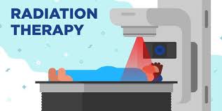
Study of the precise
application of ionizing radiation in the treatment of neoplastic growth, a
complete and effective treatment plan as well as patient care of oncology
cases. Deals with complete understanding of the principles of Radiation
treatment and the behavior of equipment used for treatment. It is also a study
of the different effects of radiation to cancer and normal cells and an
understanding of the treatment preparation process. It presents different
modalities used in the management of different types of cancer and its effects
to the different organs of the body.
This course is a study of the various pathological conditions and its effects on radiological procedures, techniques and overall radiographic image. It also discusses the specific radiographic descriptions and appearances of different modalities used in imaging.
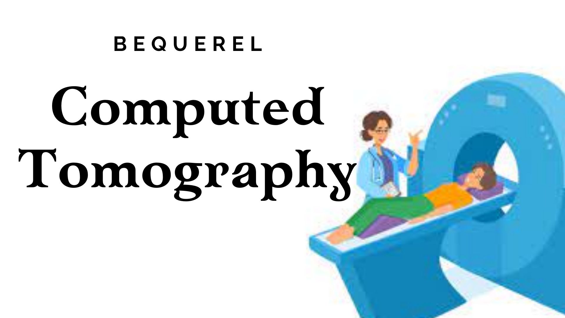
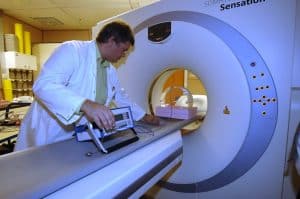
In
this course, students will be able to apply their basic knowledge about x-ray
and its application to other machines. This will help them expand their perspective
of the Radiologic Technology profession. This subject will be about the study
of the principles involved in Computed Tomography as diagnostic modality that
will demonstrate cross-sectional, transaxial, coronal and sagittal images of
the human body. It discusses the continued refinement of the current physical
principles; introduction of new principles and the development of engineering
tools to make scanners consistently perform at a level that meets the needs of
various clinical imaging requirements.
Janine Claire D. Buco, RRT - Clinical Instructor